Multimodal Analysis of Neurodegeneratie Diseases and TherapiesLaboratory of Neurodegenerative Diseases, UP Saclay CEA CNRS (UMR-9199), MIRCen, Fontenay aux Roses, FranceMarc Dhenain----------------------------------------
Alzheimer's disease: new mechanistic insights for future therapies Alzheimer's disease is the consequence of the accumulation in the brain of three lesions: amyloid-β (Aβ) and tau lesions and neuroinflammation. Our group evaluates mechanisms associated with the occurrence of these lesions and with the impact of these lesions on synaptic loss and functional impairments associated with Alzheimer's disease. > Alzheimer's disease pathology is transmissible including in primates Our group has demonstrated that Aβ and tau lesions are transmissible and can spread in the brain of mice and in primates (Gary, 2019). This transmission makes it possible to explore the pathophysiogenic mechanisms leading to the pathology. In addition to studies in mice, we investigate Alzheimer pathology in primates (Microcebus murinus). The microcebe is a model of neurodegenerative pathologies linked to aging. This animal presents, as it ages, cognitive alterations, alterations in cerebral metabolism, cerebral atrophy and amyloid deposits.
Amyloid and tau lesions induced in the brain of experimental models following inoculation of amyloid and tau nucleating factors. Examples of ongoing projects
The assembly of Aβ and tau proteins into fibrils plays an essential role in mediating the self-propagation and cell-to-cell transmission of pathological amyloid fibrils in Alzheimer's disease. We aim to characterize the heterogeneity of Aβ and Tau amyloid fibrils present in humans, mice and primates and assess the relationships between this diversity and their pathological impacts. Examples of ongoing projects
> Spreading mechanisms in Alzheimer's disease Self-replication and transcellular spread of tau proteins are responsible for the progressive accumulation of misfolded Tau protein deposits in the brain of Alzheimer’s disease patients. Tau spreading correlates with the severity of cognitive decline. Our group characterizes factors mediating and regulating tau spreading as they represent relevant targets for future therapies. 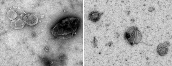
Brain-derived extracellular vesicles visualized by negative-staining electron microscopy (M. Kabani) Examples of ongoing projects
Innovative imaging tools for a better understanding of neurodegenerative diseases Neurodegenerative diseases are related to many different "small scale events" (pathological protein accumulation, neuroinflammation, cellular alterations) that lead to large-scale events (tissue loss, neuronal networks alterations, cognitive impairments). Our team develops tools to integrate events occurring at different scales. These new tools require advanced imaging skills combined with artificial intelligence, big data management and high performance computing.
Overview of the range of imaging methods implemented by our group. State of the art methods to manipulate large amounts of data. These methods rely on high performance computing (HPC). > 3D microscopic imaging methods based on high performing computing (HPC) and artificial intelligence
Our group implements image-processing pipelines to perform 3D histology in primates and rodents. 3D-reconstructed brain samples can be analyzed using semi-automatic manual analysis, digital atlas-based analysis (Lebenberg, 2011) or voxel-wise SPM approach without a priori (Vandenberghe, 2018). The method can be used to detect lesions as amyloid plaque related to Alzheimer (Vandenberghe, 2018). New methods are developed to integrate artificial intelligence, massive data management and high performance computing in our analyses. Our methods are mainly developed using in-house software platform BrainVISA (http://brainvisa.info) and high performance computing (HPC) resources (supercomputer of the TGCC - CEA, Bruyères-le-Châtel).
Quantification of neuronal density and other neuronal parameters based on high performance computing. Brain sections stained for cells (NeuN antibody) are segmented and various parameters reflecting neuronal characteristics (e.g. their density, size, orientation, etc…) are calculated. Parametric maps reflecting neuronal states at the level of the whole brain can then be produced (example in macaque brain hippocampus). Examples of ongoing projects
> In vivo imaging of functional networks Individual cells function in a harmonized way that leads to harmonious brain activity through functional networks. These networks can be assessed by resting state functional MRI and sophisticated image processing tools. Our group studies brain activity with resting state functional imaging. Our group implemented methods to detect neuronal networks in rodents (Celestine, 2020; https://sammba-mri.github.io/; Grandjean, 2020) and primates (Garin, 2021).
Example of detection of neural networks in humans and in the smallest primate in the world (mouse lemur) by magnetic resonance imaging. Examples of ongoing projects
2022: Differences between default mode networks in humans and three non-hominoid primates 2022: Automatic detection of individual neurons from the whole brain by artificial intelligence 2022: First characterization of an in vivo glutamate marker by MR imaging in a primate 2021: Tau pathology is transmissible in primates 2020: Amyloid pathology can be transmitted from furtive forms of amyloid-beta 2019: Amyloid pathology is transmissible in primates 2020: Creation of a software to analyze resting-state fMRI dataset in small animals 2018: Lifespan can be strongly increased in primates thanks to caloric restriction 2016: Reference method for 3D histology of rodent brains 2015: First demonstration that extracellular vesicles mediate prion propagation in yeast Members of the laboratory associated with these projects Luc Bousset (researcher CNRS) - https://orcid.org/0000-0002-0433-4337 Thierry Delzescaux (research director CEA, HDR) - https://orcid.org/0000-0002-6527-7946 Marc Dhenain (research director, CNRS, DVM, HDR) - https://orcid.org/0000-0001-8804-4101 Mehdi Kabani (researcher CNRS, HDR) - https://orcid.org/0000-0001-7440-6394 Jean-Luc Picq (researcher U P XIII, Professor) - https://orcid.org/0000-0003-1872-3437
Anne-Sophie Hérard (research engineer CEA) - https://orcid.org/0000-0001-8260-9618 Camille Mabillon (technician CEA, histology) Fanny Petit (technician CEA, histology) - https://orcid.org/0000-0003-2757-3434 Nicolas Souedet (research engineer CEA, high performance computing)
Marina Celestine (PhD student, Paris Saclay University) Lilian Mehl (France Relance) François Plumerault (France Relance) Huaqian Wu (PhD student, DIM)
External collaborations - Ana -Andreea Arteni, Stéphane Bressanelli, I2BC, Gif sur Yvette, France - Luc Buée, David Blum - Université de Lille, France - Alain Buisson - Grenoble Institute of Neurosciences, France - Gael Chételat - Caen University, France - Christos Constantinidis, Vanderbilt University, USA - Jean-Philippe Deslys, SEPIA, Fontenay aux Roses, France - Joanes Grandjean - Radboud University Medical Centre, The Netherlands - Stéphane Haïk, Benoît Delatour - Institut du Cerveau, France - Han Wei Hou - Nanyang Technological University, Singapour - Dulce Papy-Garcia, Université Paris Créteil, France - Fabien Pifféri, Fabienne Aujard - CNRS, Brunoy, France - Cyril Poupon - Neurospin, Gif sur Yvette, France - Human Rezaei, Vincent Beringue – INRAE, Jouy en Josas, France - Stephen Sawiak - Cambridge University, UK - Elisabeth Traiffort – INSERM, Le Kremlin-Bicêtre, France - Ina Vorberg – DZNE, Bonn, Allemagne Industrial partnersWitsee - https://www.witsee.ai/academic/ Imagine Optic - https://www.imagine-optic.com/ ANR 2020-2024. PrionDiff Impact of replication and structural diversification of prions on their cerebral dissemination ANR 2020-2023. SUMMIT: Small vessel diseases: Ultrastructure & microvasculature computational model to refine individual treatment France Alzheimer 2022-2023: Impact of specific amyloid strains on Alzheimer’s disease progression: Towards the understanding of the pivotal role of amyloid/Tau interaction on and spreading of Alzheimer’s disease mechanisms on the pathology Graduate School LSH – Paris Saclay University 2022. CryoMET. Exploiting flow cytometry methods for ex-vivo purification of extra-large complexes for cryo-EM structure analysis FRISBI 2021&2022: Characterization of brain-derived and extracellular vesicles-associated Tau seeds at the origin of Alzheimer’s disease Ministère de l'Enseignement Supérieur, de la Recherche et de l'Innovation-2022-2023. Impact of specific amyloid strains on Alzheimer’s disease progression: Towards the understanding of the pivotal role of amyloid/Tau interaction on and spreading of Alzheimer’s disease mechanisms on the pathology. PTC-SN 2021-2022. Modèles computationnels pour le décodage in-vivo de la cytoarchitecture du cortex cérébral humain Service santé des Armées . Activation des processus inflammatoires périphériques et centraux à la suite d’un traumatisme crânien : implication des systèmes catécholaminergique et corticotrope Région IdF 2019-2022. CARTOBRAIN - Cartographie haute résolution des réseaux de neurones par microscopie optique en recherche préclinique DIM Elicit 2020-2023. ENCOMPASS - Evaluation of neuronal counting methods in preclinical studies DARI-A10-TGCC. Calcul intensif pour l'analyse par machine learning de données massives 3D de microscopie de cerveaux France Relance: image processing developments to improve lightsheet images analysis Alumni
2022 Garin C. M., Hori Y., Everling S., Whitlow C. T., Calabro F. J., Luna B., Froesel M., Gacoin M., Ben Hamed S, Dhenain M., Constantinidis C. An evolutionary gap in primate default mode network organization. Cell Reports. 39, 2, 110669, April 12, 2022. https://doi.org/10.1016/j.celrep.2022.110669 You Z, Jiang M, Shi Z, Shi C, Du S, Liang J, Hérard AS, Jan C, Souedet N, Delzescaux T. Macaque Neuron instance segmentation only with point annotations based on multiscale fully convolutional regression neural network. Neural Computing and Applications. 2021. 34, 2925–2938 (2022). https://doi.org/10.1007/s00521-021-06574-7 Garin C. M., Hori Y., Everling S., Whitlow C. T., Calabro F. J., Luna B., Froesel M., Gacoin M., Ben Hamed S, Dhenain M., Constantinidis C. An evolutionary gap in primate default mode network organization. Cell Reports. 39, 2, 110669, April 12, 2022. https://doi.org/10.1016/j.celrep.2022.110669 Garin C. M., Nadkarni N. A., Pépin J., Flament J., Dhenain M. Whole brain mapping of glutamate distribution in adult and old primates at 11.7 T. 2022. NeuroImage, 251, 1 May 2022, article. 118984. https://doi.org/10.1016/j.neuroimage.2022.118984 De Giorgi F, Abdul-Shukkoor MB, Kashyrina M, Largitte LA, De Nuccio F, Kauffmann B, Lends A, Laferrière F, Bonhommeau S, Lofrumento DD, Bousset L, Bezard E, Buffeteau T, Loquet A, Ichas F. Neurons with Cat's Eyes: A synthetic strain of α-synuclein fibrils seeding neuronal intranuclear inclusions. Biomolecules. 2022 Mar 11. https://doi.org/10.3390/biom12030436 - Sébastien Piluso (2018-2022) : Automatic segmentation of individual sections of mouse brains by 3D digital atlas. PhD Thesis 2021 You, Z., M. Jiang, Z. Shi, X. Ning, C. Shi, S. Du, A. S. Hérard, C. Jan, N. Souedet, Delzescaux T.. Evaluation of automated segmentation algorithms for neurons in macaque cerebral microscopic images. Microscopy Research and Technique. 2021: 27 Apr 2021: https://doi.org/2010.1002/jemt.23786. Lam S, Petit F, Hérard A-S, Boluda S, Eddarkaoui S, Guillermier M, et al. Transmission of amyloid-beta and tau pathologies is associated with cognitive impairments in a primate. Acta Neuropathol Commun 2021; 9: 165. https://doi.org/10.1186/s40478-021-01266-8. Lam S., Boluda S., Hérard A.S., Petit F., Eddarkaoui S., Cambon K., The Brainbank Neuro-CEB Neuropathology Network, Picq J.L., Buée L., Duyckaerts C., Haïk S., Dhenain M. Alzheimer’s brain inoculation in Aß-plaque bearing mice: synaptic loss is linked to tau seeding and low microglial activity. BioRxiv. 2021. https://doi.org/10.1101/2021.04.06.438654 Kabani M.. Extracellular Vesicles-encapsulated yeast prions and what they can tell us about the physical nature of propagons. International Journal of Molecular Sciences, MDPI, 2020, 22 (1), pp.90. https://doi.org/10.3390/ijms22010090 Garin C. M., Nadkarni N. A., Landeau B., Chételat G., Picq J-L, Bougacha S., Dhenain M. Resting state functional atlas and cerebral networks in mouse lemur primates at 11.7 Tesla. NeuroImage, 226, 117589, 2021. https://doi.org/10.1016/j.neuroimage.2020.117589 Suzanne Lam (2017-2021) : "Transmission of Alzheimer pathology in murine and primate models: from proteinopathies to neuronal and cognitive impairments". PhD Thesis 2020 Herard A.S., Petit F., Gary C., Guillermier M., Boluda S., Garin C. M., French Neuropathology Network, Lam S., Dhenain M.. Induction of amyloid-beta deposits from serially transmitted, histologically silent, A-beta seeds issued from human brains. Acta Neuropathologica Communications. 8, Article number: 205, 2020. https://doi.org/10.1186/s40478-020-01081-7 Dhenain M. Modèles primates et innovations thérapeutiques contre les maladies du système nerveux central. La Lettre de l'Académie des Sciences. 2020. n°40, p34. 2020 - https://www.academie-sciences.fr/pdf/lettre/lettre40.pdf. Li T., Martin E., Abada Y., Boucher C., Cès A., Youssef I., Fenaux G., Forand Y., Legrand A., Nadkarni N., Dhenain M., Hermine O., Dubreuil P., Delarasse C., Delatour B. Effects of chronic masitinib treatment in APPPS1dE9 transgenic mice modeling Alzheimer's disease, 2020. Journal of Alzheimer's Disease. 76.4. 1339-1345, 2020. https://doi.org/10.3233/JAD-200466 Celestine M.*, Nadkarni N.A.*, Garin C., Bougacha S.*, Dhenain M. Sammba-MRI, a library for small animal neuroimaging data processing in Python. Frontiers in NeuroInformatics. 28 May 2020 | https://doi.org/10.3389/fninf.2020.00024. (These three authors participated equally to the work) Ioanas H-I., Marks M., Garin C. M., Dhenain M., Yanik M. F., Rudin M.. An automated open-source workflow for standards-compliant integration of small animal magnetic resonance imaging data. Frontiers in Neuroinformatics. 2020. https://doi.org/10.3389/fninf.2020.00005 Mandino F., Cerri D. H., Garin C. M., Straathof M., van Tilborg G. A. F., Chakravarty M. M., Dhenain M., Dijkhuizen R. M., Gozzi A., Hess A., Keilholz S. D., Lerch J. P., Ian Shih Y-Y., Grandjean J. Animal functional magnetic resonance imaging: Trends and path toward standardization. Frontiers in Neuroinformatics. 2020. Vol. 13. Art 78. https://doi.org/10.3389/fninf.2019.00078. Grandjean J., Canella C., Anckaerts C., Ayrancı G, Bougacha S., Bienert T., Buehlmann D., Coletta L., Gallino D., Gass N., Garin C. M. , Nadkarni N. A. , Hübner N., Karatas M., Komaki Y., Kreitz S., Mandino F., Mechling A. E., Sato C., Sauer K., Shah D., Strobelt S., Takata N., Wank I., Wu T., Yahata N., Yun Yeow L., Yee Y., Aoki I. , Chakravarty M. M., Chang W-T., Dhenain M., Von Elverfeldt D., Harsan L. A., Hess A., Jiang T., Keliris G. A., Lerch J. P., Okano H., Rudin M., Sartorius A., Van der Linden A, Verhoye M., Weber-Fahr W., Wenderoth N., Zerbi V., Gozzi A. Common functional networks in the mouse brain revealed by multi-centre resting-state fMRI analysis. NeuroImage. 2020. 205, Article 116278. https://doi.org/10.1016/j.neuroimage.2019.116278 Dhenain Marc. Estimation of COVID-19 cases in France and in different countries: Homogeneisation based on mortality. MedRxiv. https://doi.org/10.1101/2020.04.07.20055913 2019 Gary C., Lam S.*, Herard A.S.*, Koch J.E., Petit F., Gipchtein P., Sawiak S.J., Caillierez R., Eddarkaoui S., Colin M., Aujard F., Deslys J.P., French Neuropathology Network, Brouillet E., Buée L., Comoy E.E., Pifferi F.*, Picq J-L*, Dhenain M., Encephalopathy induced by Alzheimer brain inoculation in a non-human primate. Acta Neuropathologica Communications. 2019. 7: 126. https://doi.org/10.1186/s40478-019-0771-x Pifferi F., Terrien J., Perret M., Epelbaum J., Blanc S., Picq J.L.*, Dhenain M.*, Aujard F.. Promoting healthspan and lifespan with caloric restriction in primates. Communication Biology, Nature Publishing Group. 2019. 2, 107. 7 https://doi.org/10.1038/s42003-019-0348-z Nadkarni N. A, Bougacha S., Garin C., Dhenain M., Picq J.-L.. A 3D population-based brain atlas of the mouse lemur primate with examples of applications in aging studies and comparative anatomy. NeuroImage. 2019. 185. 85-95. https://doi.org/10.1016/j.neuroimage.2018.10.010 Levy J., Facchinetti P., Jan C., Achour M., Bouvier C., Brunet J.F., Delzescaux T., Giuliano F. Tridimensional mapping of Phox2b expressing neurons in the brainstem of adult Macaca fascicularis and identification of the retrotrapezoid nucleus. Journal of Comparative Neurology. 2019. May 9. 1-10. https://doi.org/10.1002/cne.24713 You Z, Balbastre Y, Bouvier C., Souedet N., Gipchtein P, Hantraye P, Jan C, Herard A-S, Delzescaux T.. Automated individualization of size-varying neurons in 2D microscopic images of macaque brain. 2019. Front Neuroanat. Dec 17;13:98. https://doi.org/10.3389/fnana.2019.00098 Clément Garin (2015-2019): "Characterization of mouse lemur brain by anatomical, functional and glutamate MRI". PhD Thesis. Autres Vandenberghe, M.E., Souedet, N., Herard, A.S., Ayral, A.M., Letronne, F., Balbastre, Y., Sadouni, E., Hantraye, P., Dhenain, M., Frouin, F., Lambert, J.C., Delzescaux, T. Voxel-based statistical analysis of 3D immunostained tissue imaging. Frontiers in Neuroscience. 2018; 12(article number 754). https://doi.org/10.3389/fnins.2018.00754 Lebenberg, J., Herard, A.S., Dubois, A., Dhenain, M., Hantraye, P., Delzescaux, T. A combination of atlas-based and voxel-wise approaches to analyze metabolic changes in autoradiographic data from Alzheimer's mice. Neuroimage. 2011. 57(4): 1447-1457. https://doi.org/10.1016/j.neuroimage.2011.04.059 Organigramme of the Laboratory of Neurodegenerative Diseases
|
|||||||||||||
|---|---|---|---|---|---|---|---|---|---|---|---|---|---|